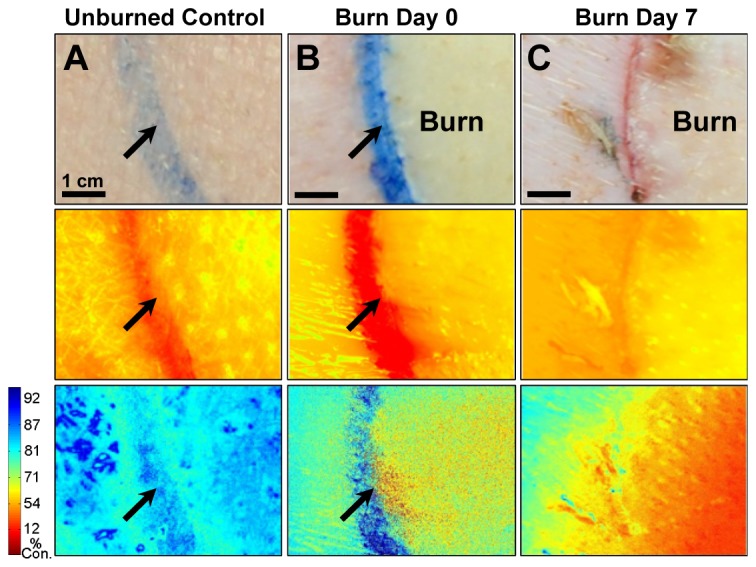
Fig. 5.

Progression of a full-thickness burn on the paraspinal skin of a Yorkshire pig monitored by oxygen-sensing paint-on bandage. Images shown for A) control skin; B) immediately post-burn and C) 7 days post-burn. Top row: regular photographs; middle row: emission at 700 nm - oxygen-dependent phosphorescence signals are overwhelmed by the skin autofluorescence background; bottom-row: percent oxygen consumption (%) maps obtained after eliminating autofluorescence - blue indicates higher oxygen consumption by tissues. Red indicates lower oxygen consumption by tissues. Arrows indicate ink marks dividing burn and surrounding skin.
