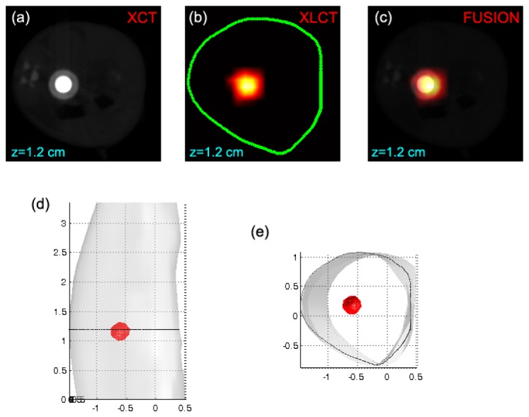
Fig. 4.

The in vivo XLCT reconstruction results for illustrating the performance of the proposed method. In the experiment, a transparent tube filled with the NIR-emitting nanophosphor (Gd2O3:Eu3+) was implanted into the body of the mouse. (a) The reconstructed XCT tomographic image. (b) The reconstructed XLCT tomographic image. These XLCT tomographic images were obtained by the use of the proposed wavelet-based reconstruction method with wavelet components retained. In addition, only single-view data [see Fig. 3(a)] was used in the XLCT reconstruction. The green curve in (b) depict the mouse boundary obtained by back-projecting the 72 white light images. This method is similar to that described in [25]. (c) The fusion image of the XLCT and XCT images. (d) and (e) The 3-D visualization results of the reconstructed XLCT tomographic images from two views. The black circles in (d) and (e) indicate the positions of investigated slice.
