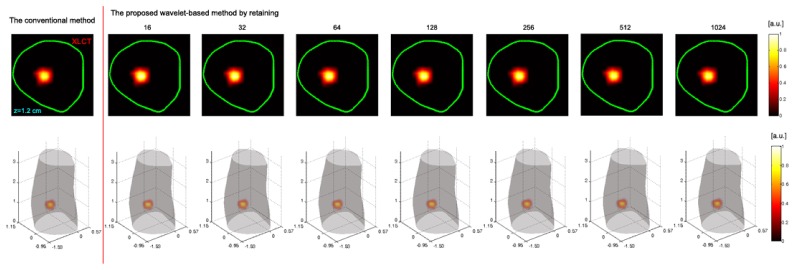
Fig. 5.

Comparison of the reconstruction results in the in vivo experiment, obtained by the conventional method (i.e., using the non-reduced weight matrix) and the proposed method (i.e., using the reduced weight matrix) with different components retained. The 1st column shows the reconstruction results obtained by the conventional method. In the conventional reconstruction, the weight matrix was generated from 11215 measurements based on Eq. (4). The 2nd-8th columns show the reconstruction results obtained by the proposed wavelet-based method with 16, 32, 64, 128, 256, 512, and 1024 components retained, respectively. The top row shows the reconstructed 2-D XLCT tomographic images. The bottom row shows the corresponding the 3-D visualization results. The reconstructed images in 1st and 2nd rows are normalized by the maximum of the reconstructed results and then displayed on the same color scale, respectively.
