
Fig. 2.
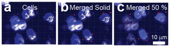
Gold nanostars are located inside and around nuclei of fixed cerebellar neurons. (a-c) Volumetric reconstruction of the molecular layer of a cerebellar slice incubated with gold nanostars. (a) Reconstruction of the nucleus of 10 molecular layer neurons stained with DAPI. Images obtained from the channel filtered between 435 to 485 nm. (b) Identification of nanoparticle clusters (red dots) close to the nuclei. Image obtained by merging volumetric data from the two channels. (c) Same data as in (b) with the transparency of the reconstructed surfaces set to 50% showing the presence of nanoparticles inside the nuclei.