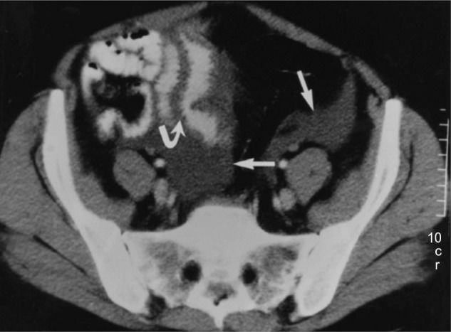
Figure 4.

Abdominal computed tomography scan of patient with hereditary angioedema showing thickening of the small bowel (stacked-coin appearance) due to angioedema.
Notes: Curved arrow indicates prominent fold thickening; straight arrows indicate pelvic ascites. Reprinted with permission from the American Journal of Roentgenology. De Backer AI, De Schepper AM, Vandevenne JE, Schoeters P, Michielsen P, Stevens WJ. CT of angioedema of the small bowel. Am J Roentgenol. 2001;176(3):649–652.39
