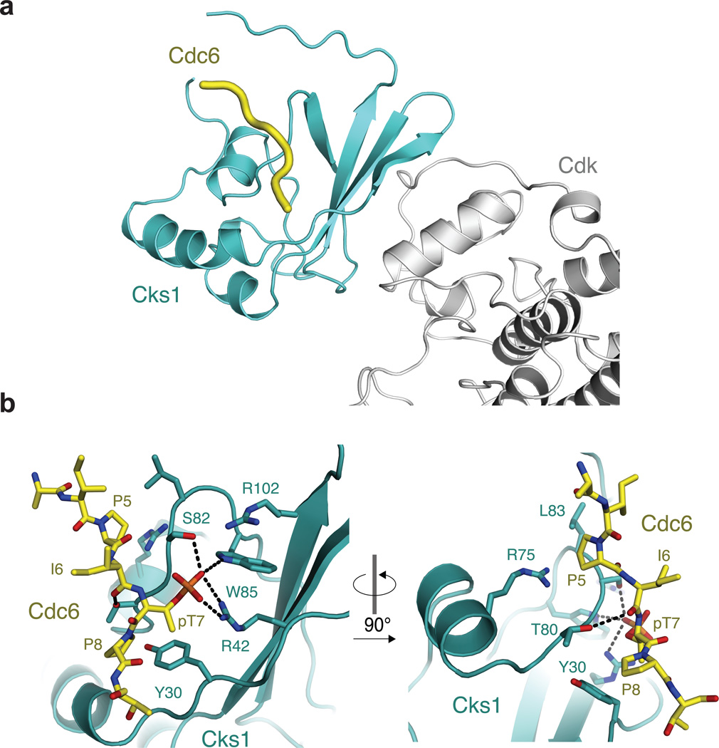
Figure 3.
Crystal structure of a phosCdc63–9 -Cks11–112 complex. (a) Ternary complex including Cdk shows that the phosphorylated substrate binding site in Cks is distal from Cdk. The model was created by aligning Cks1 from the structure solved here and hsCks1 in the hsCks1-Cdk2 complex (PDB code: 1buh35). (b) Close-up views of the phosCdc63–9 binding site in Cks1.

