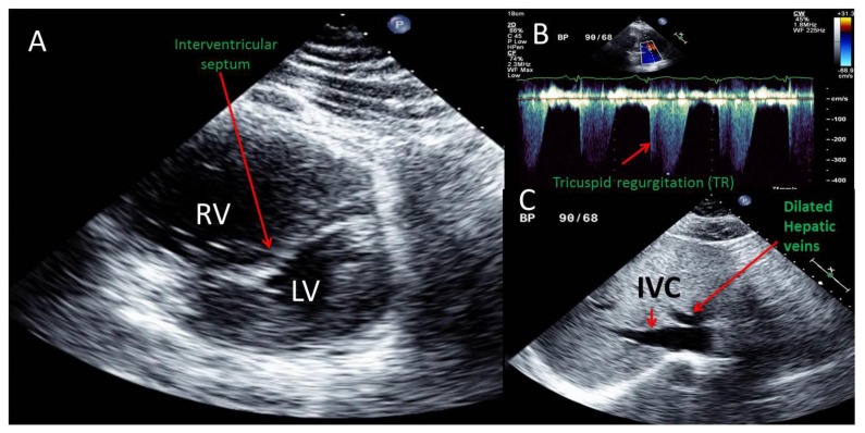
Figure 3.
A 51 year old male with saddle pulmonary embolus and impending paradoxical embolism. Transthoracic echocardiographic (TTE) images are shown. Findings: A. Parasternal short axis view showing a severely dilated right ventricle (RV) severely compressing and deforming the left ventricle (LV) into a D shape due to marked deformity of the interventricular septum during late systolic and throughout diastole indicative of severe right ventricular pressure and volume overload.
B. Continuous wave Doppler of tricuspid regurgitation obtained from an off axis apical view demonstrating tricuspid regurgitation (TR) with a peak velocity 3.7 m/sec which using the simplified Bernoulli equation would correspond to an RV systolic pressure of 54 mmHg. Note that TR peak velocity is higher with every other beat and represent an equivalent of pulsus alternans due to severe RV systolic dysfunction. With the addition of an estimated right atrial pressure of 15–20 mmHg, the estimated pulmonary artery systolic pressure was 70–75 mmHg.
C. Subcostal view illustrating a plethoric inferior vena cava (IVC) with minimal collapse to inspiration indicating an estimated right atrial pressure of 15–20 mmHg. Also, note plethora of the hepatic veins also consistent with significantly elevated right atrial pressure. Technique: IE_33 Philips echocardiography machine, 2 MHz transducer

