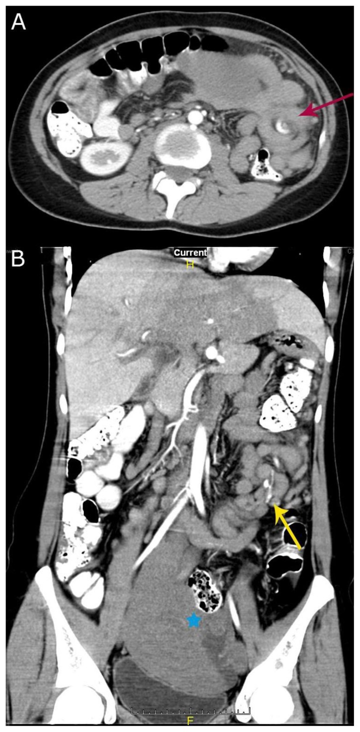
Figure 4.
29 yr old female patient with ectopic, torsed & infarcted spleen. (a). Axial post contrast CT scan in the arterial phase shows classic whorled appearance of twisted splenic vascular pedicle (red arrow) in the axial image, containing engorged non-enhanced splenic vein and enhanced splenic artery. (b) Coronal reconstructed CT image in the arterial phase shows multiple (five) twists of the splenic vascular pedicle, the splenic hilum (blue asterix) is facing left laterally.
(Siemens SOMATOM Definition Flash, kVp 120, mA 772, 5 mm slice thickness, Pitch 1.375:1, 100 cc of Visipaque 320 IV, 900ml diluted gastrograffin Orally, in arterial phase)

