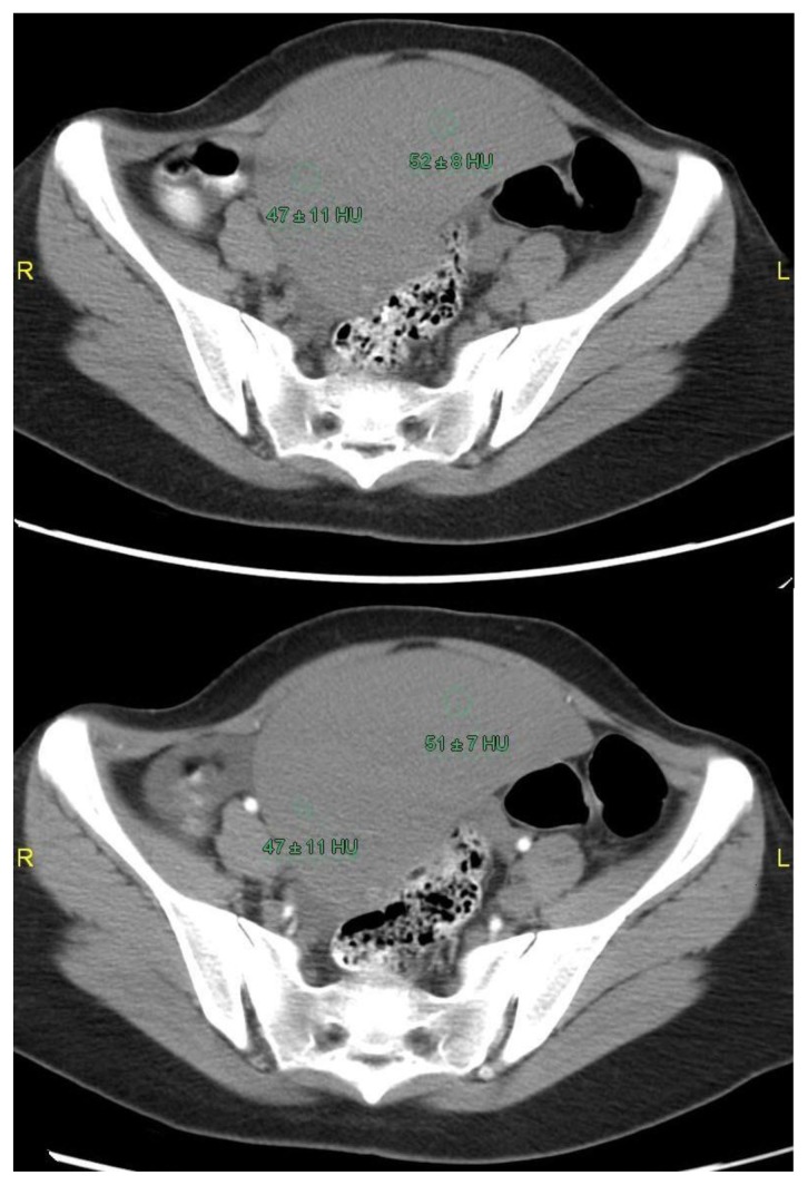
Figure 7.
29 yr old female patient with ectopic, torsed & infarcted spleen. (a) Pre and (b) post contrast axial CT images show no definite change in the attenuation of the splenic parenchyma denoting complete infarction. Enhancing rim of the splenic capsule is seen.
(Siemens SOMATOM Definition Flash, kVp 120, mA 772, 5 mm slice thickness, Pitch 1.375:1, 100 cc of Visipaque 320 IV, 900ml diluted gastrograffin Orally, in precontrast & arterial phase)

