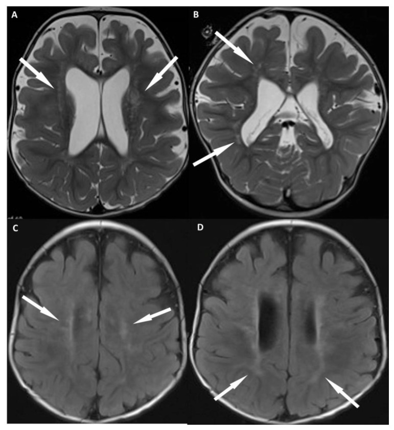
Figure 4.
12 month old with male failure to thrive and diagnosis of OCRL. FINDINGS: MR imaging of the brain shows characteristic imaging features of OCRL. Axial T2 (a) and coronal T2 (b) weighted images show prominent perivascular spaces within the periventricular and deep white matter (arrows). Axial FLAIR (c, d) show confluent regions of T2 prolongation within the periventricular white matter with sparing of the subcortical white matter (arrows), which is typical early in the course of the disease. Also, note the lack of small cystic structures in the periventricular and deep white matter, which typically don’t develop until later in the disease course. TECHNIQUE: (4a, b) Siemens 3T SKYRA MR scanner, axial T2-weighted images, TR= 3312, TE = 89, 4 mm slice thickness, no contrast. (4c, d) Siemens 3T SKYRA MR scanner, axial FLAIR images, TR= 7602 TE =132, 4 mm slice thickness, no contrast.

