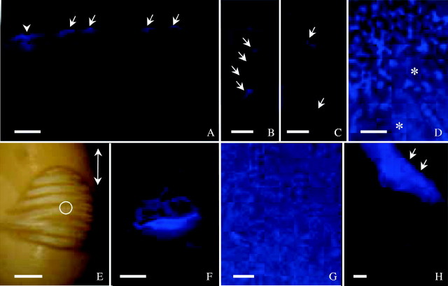
Fig. 3.
Seeds incubated in Cellufluor and observed under UV light. (A) ‘Harovinton’ seed in dye for 1 min and section made from abaxial side. Surface deposits stained (arrows), along with palisade walls subtending a crack (arrowhead). (B) and (C) ‘Tachanagaha’ seed 2 min in dye. Single cells (B) or groups of cells (C) involved in crack formation as demonstrated with the dye (arrows). (D) ‘Tachanagaha’ seed 2 min in dye, with surface deposits (Type II) stained. Areas free of large deposits were slightly stained in the outer periclinal walls of palisade layer (asterisks); this makes it difficult to detect initial sites of dye entry. (E) Initial water uptake at dorsal side of ‘OX 951’ seed after 5·5 h in dye, with wrinkles formed as water moved in. White light micrograph from a dissecting microscope. Double-headed arrow indicates the seed's long axis. A crack in the centre of the wrinkled area is circled. (F) Crack in (E) observed with UV light, illustrating intensely stained palisade walls. (G) ‘Harovinton’ seed in dye for 1 min. Heavy surface deposits at abaxial side stained. (H) ‘Harovinton’ seed in dye for 3 min, with endocarp tissue (Type III deposits, arrows) intensely stained; other deposits (Type II, lower half of micrograph) stained less intensely. Scale bars: E = 1 mm; all others = 100 µm.

