Fig. 6.

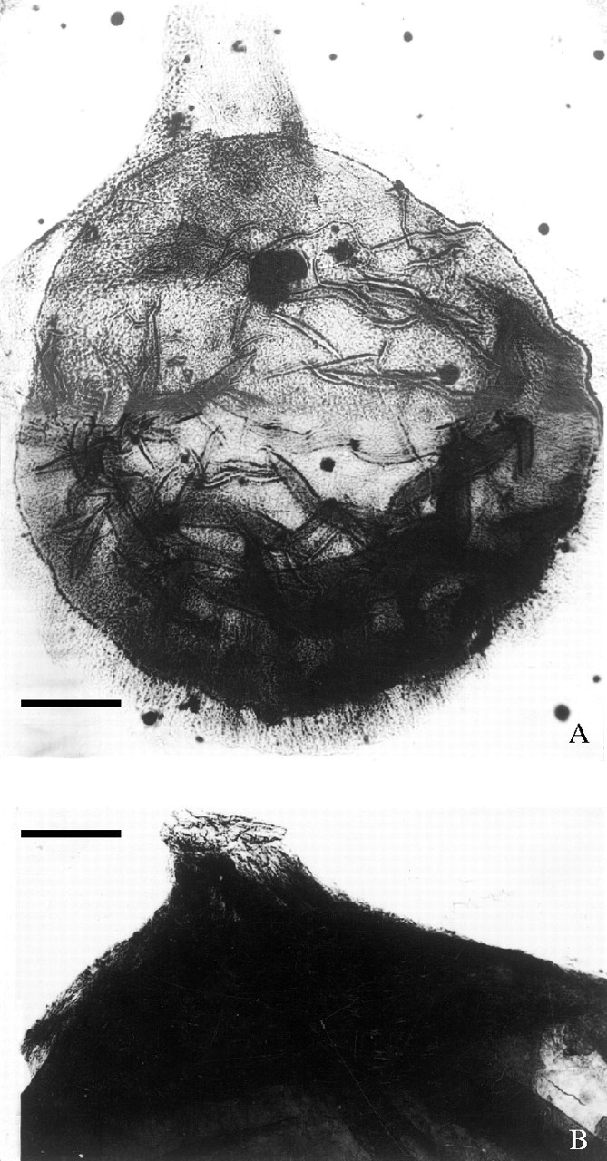
Ovules through maceration: (A) an ovule showing a long, unbifid micropylar tube at apex, 9705–0; ×40; (B) upper part of an ovule showing a prominent micropyle with an oblique tip, 9107–8, ×40. Both scale bars = 0·25 mm.

Ovules through maceration: (A) an ovule showing a long, unbifid micropylar tube at apex, 9705–0; ×40; (B) upper part of an ovule showing a prominent micropyle with an oblique tip, 9107–8, ×40. Both scale bars = 0·25 mm.