
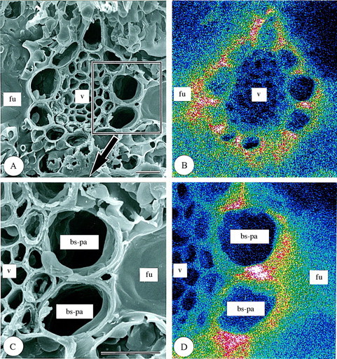
Fig. 3. Scanning electron micrographs of transverse sections of freeze‐dried first‐year leaves of Sasa veitchii. Bar = 10 µm. (A and C) Secondary electron images; (B and D) silicon distribution images by SEM–EDX. (A and B) Vascular bundle tissue; (C and D) bundle sheath parenchyma cells. bs‐pa, Bundle sheath parenchyma cell; fu, fusoid cell; v, vascular bundle tissue.