
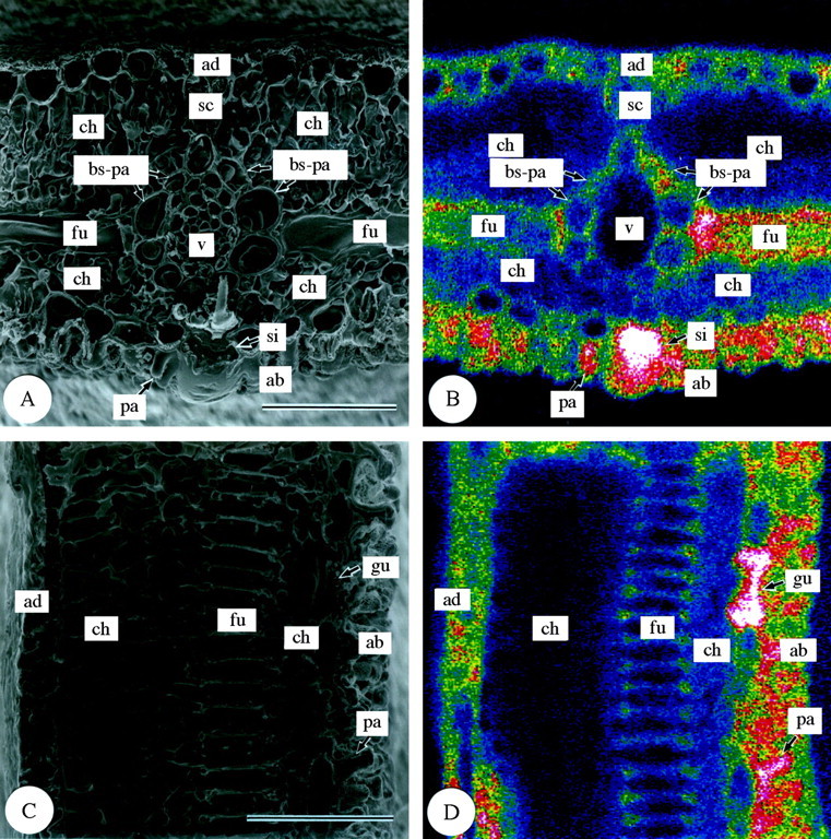
Fig. 4. Scanning electron micrographs of transverse and longitudinal sections of freeze‐dried third‐year leaves of Sasa veitchii. Bar = 50 µm. (A and C) Secondary electron images; (B and D) silicon distribution images by SEM‐EDX. (A and B) Transverse fractures; (C and D) longitudinal fractures. ad, Adaxial side; ab, abaxial side; bs‐pa, bundle sheath parenchyma cell; ch, chlorenchyma cell; fu, fusoid cell; gu, guard cell; pa, papillae; sc, sclerenchyma cell; si, silica cell; v, vascular bundle tissue.