
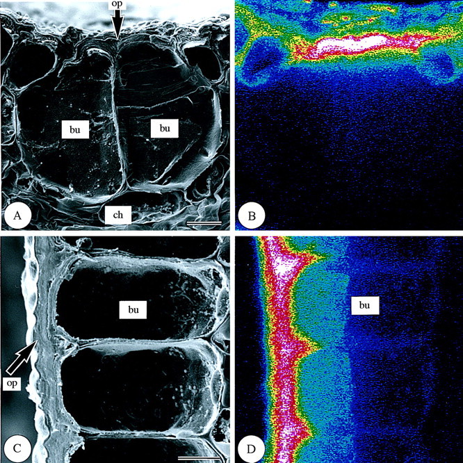
Fig. 6. Scanning electron micrographs of bulliform cells in transverse and longitudinal sections of freeze‐dried first‐year leaves of Sasa veitchii. Bar = 10 µm. (A and C) Secondary electron images; (B and D) silicon distribution images by SEM‐EDX. (A and B) Transverse fractures; (C and D) longitudinal fractures. bu, Bulliform cell; ch, chlorenchyma; op, outer periclinal cell wall.