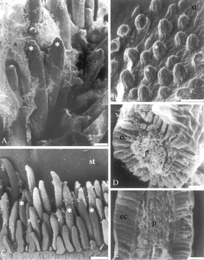
Fig. 2.

Scanning electron microscopy. (A and B) An overview of the colleters of Simira glaziovii at (A) entirely developed and (B) initial stage; (C) an overview of the colleters of S. rubra; (D) cross-section of a colleter of S. glaziovii; and (E) longitudinal section of a colleter of S. pikia. Scale bars: A and B = 100 µm; C = 250 µm; D and E = 25 µm. Asterisks, colleter; stars, immature colleters; ec, epidermal cells; p, parenchyma; s, secretion; st, stipule.
