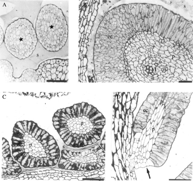
Fig. 3.
Light microscopy. (A and B) Cross-sections of the colleters of Simira glaziovii at (A) initial stage and (B) fully developed; (C) cross-section of the colleters of S. pikia; and (D) longitudinal section of a colleter of S. rubra. Scale bars: A, B and D = 50 µm; C = 60 µm. Stars, immature colleters; arrow, constriction; ec, epidermal cells; p, parenchyma; s, secretion; st, stipule; vc, vascular trace.

