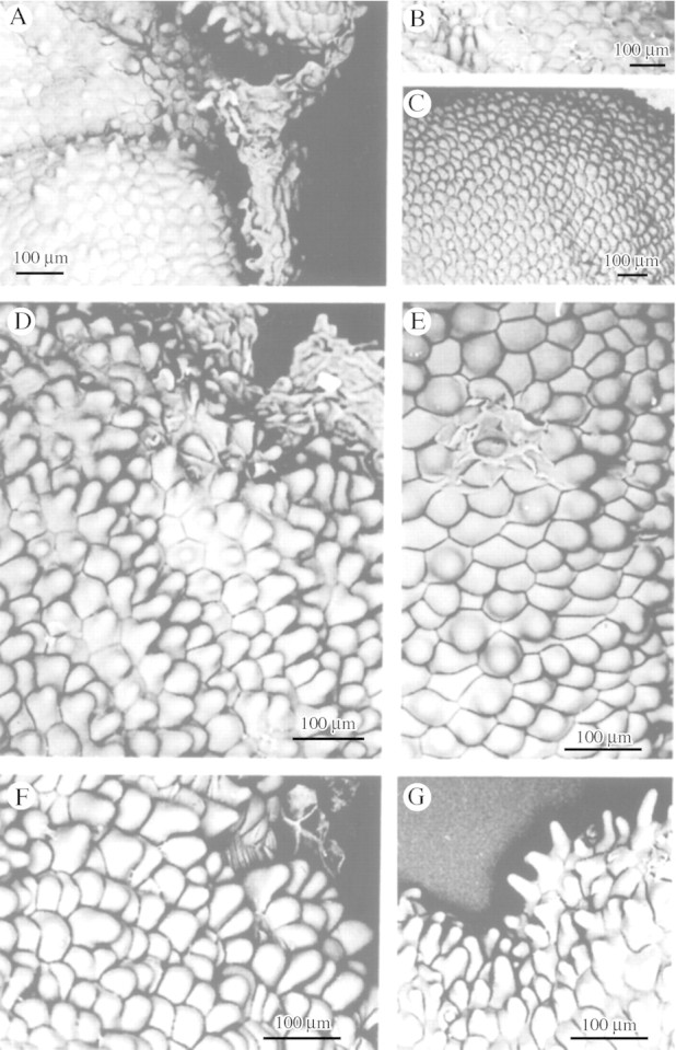
Fig. 2. Conical labellar papillae with broad points of insertion and rounded apices of M. mosenii (A), M. cogniauxiana (B), M. vernicosa (C), M. minuta (D) and M. seidelii (E). F and G, M. cf. minuta showing typical and marginal labellar papillae, respectively. Scale bar = 100 µm.
