
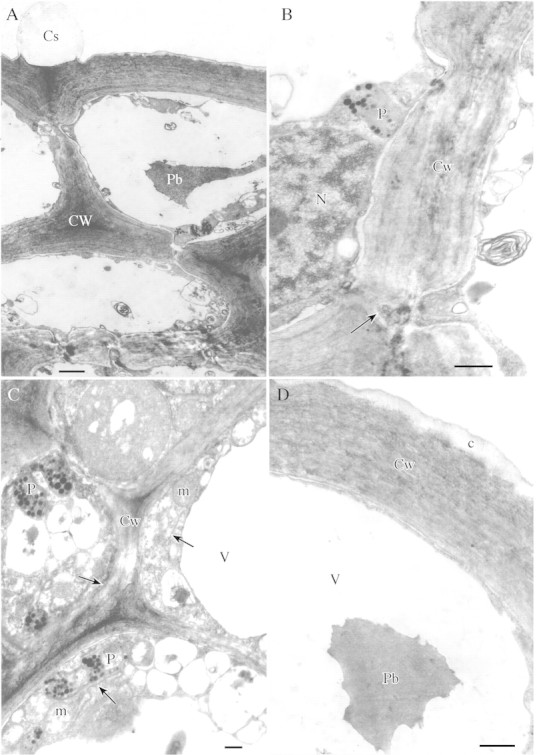
Fig. 2. A, Cell walls of secretory cells with numerous pits; the epidermal cell showing swelling of cuticle. Scale bar = 2 µm. B, Section through thick cell wall of secretory epidermis showing plasmodesma within pit (arrow). Scale bar = 1 µm. C, Cytoplasm of secretory cell with abundant mitochondria, ER (arrows) and plastids containing numerous plastoglobuli. Scale bar = 1 µm. D, Outer tangential wall of epidermis with reticulate cuticle. Note the single, finely granular, intravacuolar protein body. Scale bar = 1 µm.