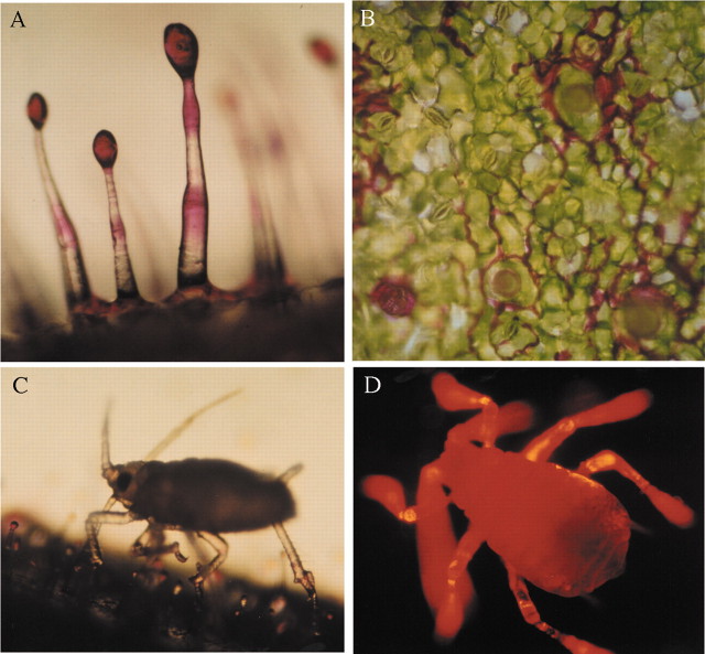
Fig. 2. A, Nicotiana tabacum GSTs after treatment with rhodamine B to stain sucrose esters of exudates. B, Top view of rhodamine B‐treated leaf showing migration of stained exudate to the base of trichomes, then out onto the epidermal surface. C, Aphid walking on a rhodamine B‐treated leaf, note exudate on the legs. D, Fluorescence micrograph of aphid after it had walked on a rhodamine B‐treated leaf for 1 min. Yellow fluorescence denotes highest stain accumulation. Control aphids show no autofluorescence.

An official website of the United States government
Here's how you know
Official websites use .gov
A
.gov website belongs to an official
government organization in the United States.
Secure .gov websites use HTTPS
A lock (
) or https:// means you've safely
connected to the .gov website. Share sensitive
information only on official, secure websites.
