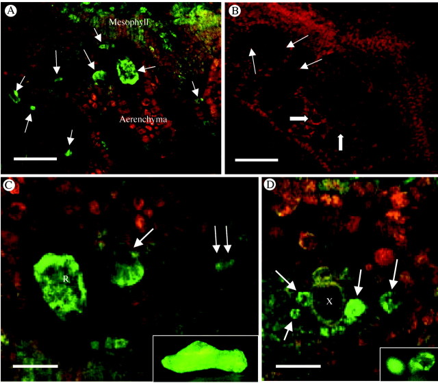
Fig. 4. Binding of green fluorescent‐tagged Ca2+ channel blocker, DM‐Bodipy‐DHP, to fresh sections of developing Pistia leaf. (A) Low magnification image. The brightest green fluorescence is associated with Ca oxalate crystal idioblasts at various stages of development (arrows). Some fluorescence is also seen in the young developing mesophyll cells. Red fluorescence is due to chlorophyll. (B) Control. Binding of DM‐Bodipy‐DHP is completely inhibited by a 4‐h pretreatment with the competitive Ca2+ channel blocker, nifedipine. Raphide crystal idioblasts (arrows) and druse idioblasts (block arrows) are pointed out for reference. (C) Enlargement from A showing labelled crystal idioblasts. The raphide idioblast to the left (R) is focused through the cortical cytoplasm indicating Ca2+ channels are abundant in cytoplasmic organelles. At the large single arrow, the same focal plane is near the surface of the raphide idioblast indicating potential association of the fluorescent probe with the plasma membrane as well. At the double arrow is a cell that is just beginning initiation of differentiation to become a raphide idioblast. Inset is a living raphide crystal idioblast stained with DiOC6 to show ER abundance. (D) Image of the xylem region of a vein where druse crystal idioblasts are most common. At least four druse idioblasts at various developmental stages are seen and are fluorescing brightly (arrows). Inset is of living druse idioblasts stained with DiOC6. R, Raphide crystal idioblast; X, xylem. Scale bars: A and B = 50 µm; C and D = 21 µm.

An official website of the United States government
Here's how you know
Official websites use .gov
A
.gov website belongs to an official
government organization in the United States.
Secure .gov websites use HTTPS
A lock (
) or https:// means you've safely
connected to the .gov website. Share sensitive
information only on official, secure websites.
