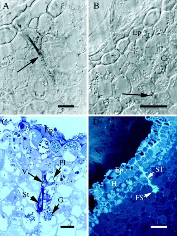
Fig. 2. Location of feeding sites in nodosities induced by isolate SRU‐1 (A–C) or VWL‐1 (D). (A) 4 µm transverse section in GMA of nodosity at point of stylet penetration (arrow), viewed with Nomarski differential interference contrast optics. Scale bar = 10 µm. (B) As for (A), adjacent section showing stylet tip (arrow). The stylet penetrates through the epidermis (Ep) and several cell layers into the cortex (C) of the root, but does not reach the stele. Scale bar = 20 µm. (C) 0·5 µm transverse section in epoxy resin, stained with TBO, showing epidermis (Ep); collapsed plasmalemma (Pl); vesicles (V); granular structures (G); section cuts obliquely through stylet (St). Scale bar = 10 µm. (D) 4 µm transverse section in GMA viewed under UV (365 nm) excitation, showing stylet track of feeding phylloxera. Epidermis (Ep); hypodermis (H); stylet track (ST); feeding site (FS). Scale bar = 50 µm.
