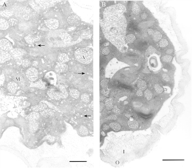
Fig. 9. (A) Pollen tube near the tip. Secretory vesicles are numerous (arrows) as are mitochondria (M). Larger Golgi vesicles are also present (V). Bar = 1 µm. (B) Transverse section of a pollen tube showing a wrinkled pectic outer layer of the cell wall (O) and loosely bound fibrils of the inner callose/cellulosic tube wall (I). Bar = 1 µm.
