
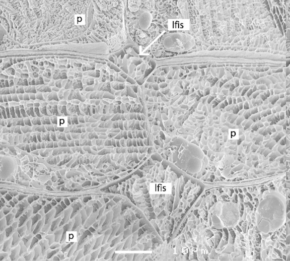
Fig. 5. Cryo‐SEM micrograph showing seed coat parenchyma cells (p) with liquid‐filled intercellular spaces (lfis). The similar mesh of the white matrix in the ice indicates a comparable solute concentration in the apoplast and in the cytosol. Bar represents 10 µm.