
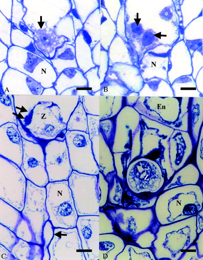
Fig. 3. Embryo sac contents at anthesis prior to pollination (A and B) and 10 d (C) and 30 d (D) post‐pollination. (A) Arrow denotes partially cellularized egg cell; (B) arrows mark partially cellularized synergids. *, Polar nucleus. (C) Single arrow marks pollen tube in nucellus; double arrow delineates penetrated synergid. Bars: A–C = 100 µm; D = 50 µm. En, Cellular endosperm; N, nucellus; Z, zygote.