
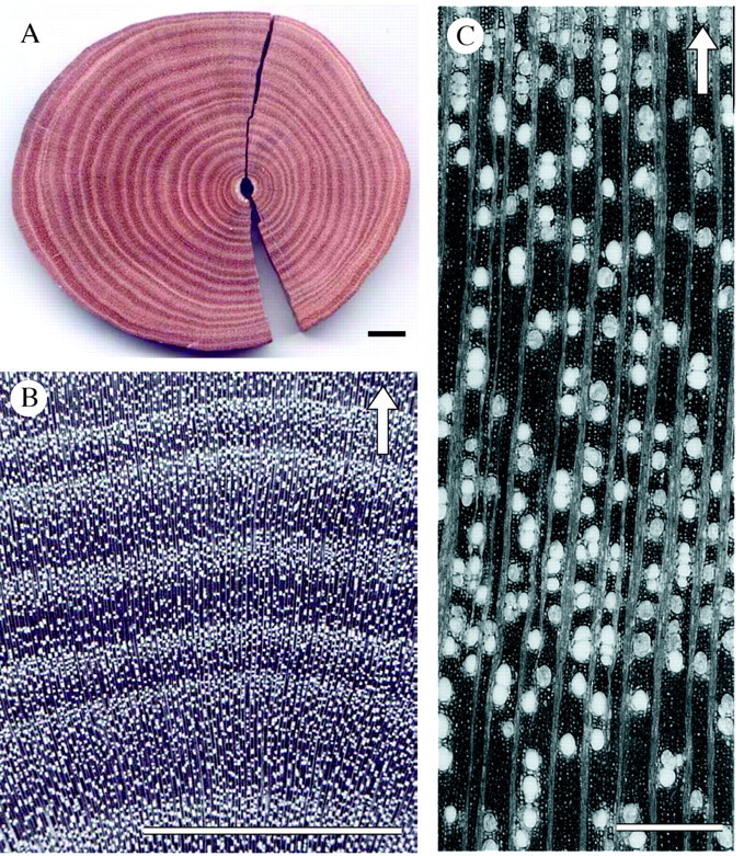
Fig. 1. (A) Macroscopic picture of a polished Rhizophora mucronata wood section showing a clear alternation of dark and light growth layers. (B) Magnified R. mucronata wood disc revealing the changing vessel density. (C) Microscopic photograph showing the gradual change in vessel density and the absence of distinct growth ring boundaries. Scale bars: A and B = 1 cm; C = 500 µm; the arrows indicate the direction of growth.