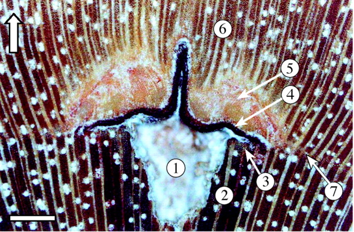
Fig. 2. Microphotograph of the cambial mark: 1, puncture canal; 2, oxidized wood; 3, fibres with incomplete cell wall thickening; 4, layer of crushed cambial derivatives; 5, parenchymatous wound tissue; 6, restored wood structure; 7, local parenchyma band indicating the position of cambial initials at the time of pinning (see text for more detailed explanation). Scale bar = 500 µm; the arrow indicates the direction of growth.
