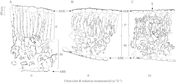
Fig. 6. Anatomical features of cotton leaves exposed to 0 (A), 8 (B) and 16 (C) kJ m–2 d–1 of ultraviolet‐B radiation. The diagrams are traced from images obtained using a Leica TCSNT Confocal laser scanning microscope attached to a pictomicrography system. The adaxial (ADE), abaxial epidermis (ABE), palisade layer (P) mesophyll layer (M) stomata (S) and intercellular cavities (IC) can be clearly seen in the diagrams. Bar = 60 µm for length and width in all panels.

An official website of the United States government
Here's how you know
Official websites use .gov
A
.gov website belongs to an official
government organization in the United States.
Secure .gov websites use HTTPS
A lock (
) or https:// means you've safely
connected to the .gov website. Share sensitive
information only on official, secure websites.
