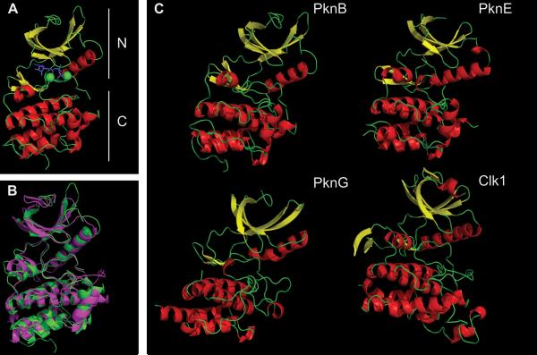
FIGURE 4.
Overview of the M. tuberculosis STPK's KD. (A) Major features of the PknB KD (1MRU_B): N-terminal (upper) and C-terminal (lower) lobes are labeled. The ATP analog is in blue, and two Mg2+ ions are in green. (B) Overlap of PknB (green) and Clk1 (magenta). Clk1 was a top hit when the PknB structure was used to search similar three-dimensional structures using the NCBI VAST program. For clarity, residues 298 to 319 and 395 to 443 in Clk1 that are absent in M. tuberculosis STPKs (see Fig. 2) are truncated in Clk1. (C) PknB (1MRU_B), PknE (2H34_B), PknG (2PZI_A), and Clk1 (1Z57). α-Helix is in red, β-sheet is in yellow. Figures were made using PyMOL (Schrödinger) and POV-Ray (povray.org). doi:10.1128/microbiolspec.MGM2-0006-2013.f4

