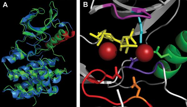
FIGURE 5.
Active site of PknB KD. (A) Overlap of “closed” PknB KD (1MRU_B) in green and “open” apo-PknE-KD (2H34) in blue, with the PknE C helix labeled in red. (B) PknB active site (1MRU_B) P loop (GFGGMS), magenta; Mg2+, red balls; ATPγS, yellow; C-helix, green (Glu59-green); Lys40, aqua; catalytic loop, red (Asp138-orange, Asn143-red); DFG motif, purple (Asp156-purple). Figures were made using PyMOL (Schrödinger) and POV-Ray (povray.org). doi:10.1128/microbiolspec.MGM2-0006-2013.f5

