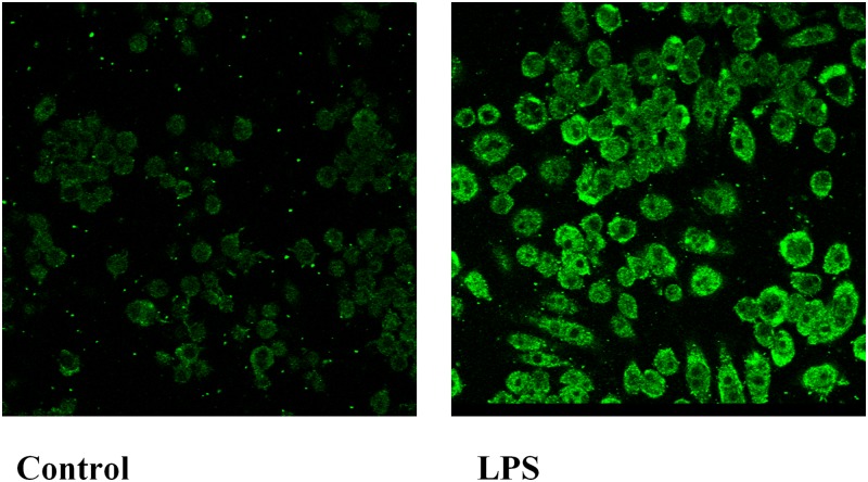
Fig. 3.
Immunofluorescence analysis for GPR109A/HCA2 in RAW cells. Cells were treated with LPS at 100 ng/ml in serum-free medium for 16 h. Immunostaining was performed as described in Materials and Methods. Fluorescent GPR109A/HCA2 staining was visualized by confocal microscopy with a 40× oil immersion objective lens. All images were acquired with identical settings.

