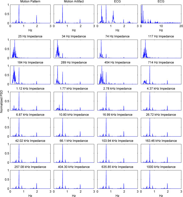
Figure 7.

PSDs of the motion pattern, motion artifact, ECG and impedance changes at each impedance current frequency. On the top row, PSDs of the motion pattern, the motion artifact, and ECG are shown from 0 Hz to 3 Hz in a normalized between 0 to 1 manner. Lastly, a normalized PSD graph of ECG is shown from 0 Hz to 20 Hz for demonstration of the signal components at frequencies higher than 3 Hz. In the following rows, the normalized PSDs of the impedance measurements are shown for each impedance current frequency. The data is filtered between 0.2 Hz and 50 Hz. The x-axes for all graphs are same, except the top right corner plot.
