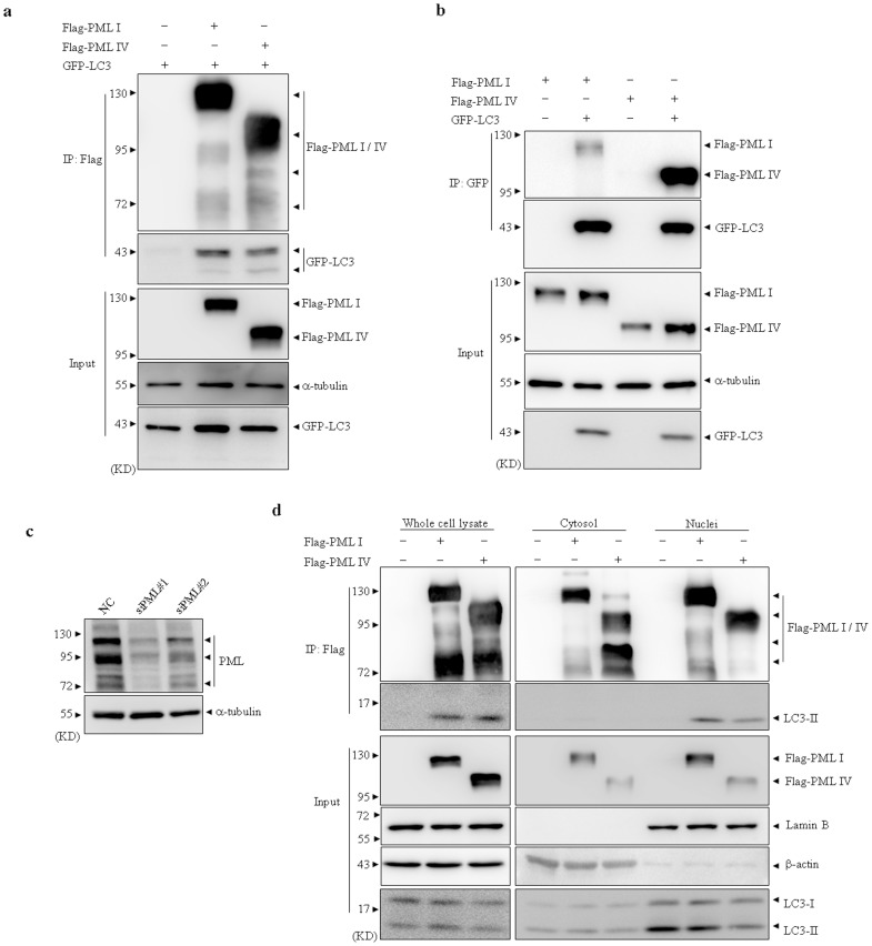
Figure 1. PML interacts with overexpressed and endogenous LC3 proteins.
(a–b) HEK293T cells were transiently transfected with the indicated plasmids. After transfection for 48 hours, whole cell lysates were harvested and Co-IP assay was performed by Flag (a) or GFP (b) antibody. Then the indicated proteins were detected by western blot. (c) HEK293T cells were stably transfected with siPML#1, siPML#2 or NC. The expression of PML protein level was detected by PML antibody with α-tubulin as loading control. (d) siPML#1-expressing HEK293T cells were transiently transfected with Flag tagged shRNA-resistant PML I, PML IV or empty vectors, then fractionated cytosol and nuclei together with whole cell lysates were applied to IP by Flag antibody. Endogenous LC3 protein was detected in immunoprecipitate by western blot. 10% cell lysates (input) was used as a positive control. All experiments were repeated for three times and similar results were obtained.

