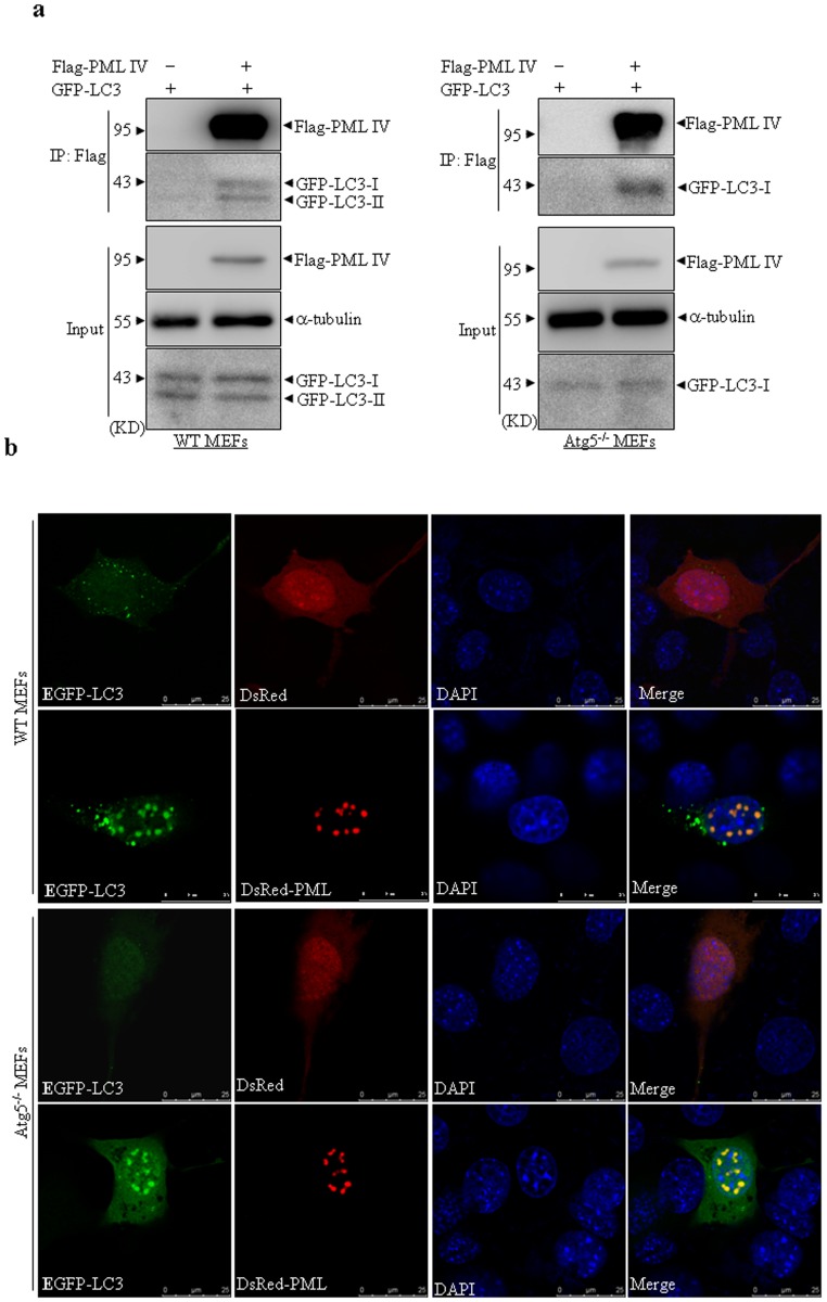
Figure 5. Interaction of PML and LC3 is independent of autophagic activity.
(a) Wild type (WT) and Atg5−/− MEF cells were respectively transfected with the indicated plasmids. After transfection for 48 hours, whole cell lysates were harvested and immunoprecipitated with Flag antibody, followed by immunoblots with GFP antibody. 10% cell lysates (input) was used as a positive control. (b) Indicated cells were co-transfected with EGFP-LC3 and DsRed-PML IV or DsRed vector, respectively. Following transfection for 24 hours, the cells were fixed and observed under confocal microscopy. Representative colocalization images of EGFP-LC3 and DsRed-PML in indicated cells were shown (scale bar = 25 µm). All experiments were repeated for three times and similar results were seen.

