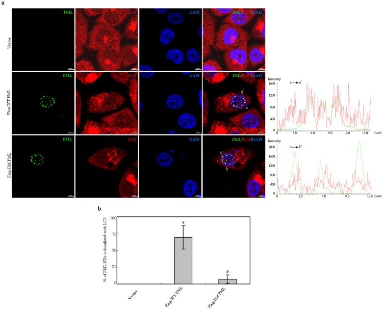
Figure 7. Effects of wild type and double mutant PML on localization of endogenous LC3 protein.
PC3 cells were transfected with Flag tagged WT and double mutant (DM) PML expressing plasmids. After transfection for 48 hours, the localization of PML and LC3 proteins were analyzed with Flag and LC3 antibodies. (a) Representative images were captured by confocal microscope (scale bars = 10 µM). Line scan analysis right was applied to quantify colocalization of LC3 and Flag tagged WT PML or DM PML crossing PML NBs as indicated on left merged images. (b) Quantification of percentages of PML NBs colocalized with LC3 per cell in part (a) was presented. Data presents mean percentage with bar as S.D by analyzing 30 cells in an independent experiment. The symbols * and # indicate p<0.01 compared with the cells expressing empty or Flag-WT PML plasmids, respectively. All experiments were repeated for three times and similar results were obtained.

