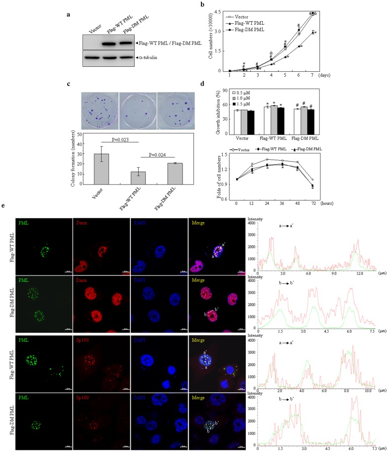
Figure 8. Effects of wild type and double mutant PML on growth and doxorubicin-induced cytotoxic activity of HEK293T cells.
(a) HEK293T cells were stably transfected with indicated plasmids. The expressions of Flag tagged WT and DM PML proteins were detected with Flag antibody. (b) Indicated cells were respectively cultured for days as indicated and followed by CCK-8 assay. (c) Dense foci formation on a monolayer of indicated cells for 15 days was observed by light microscope (upper part) and foci numbers were counted. Data represents means with bar as S.D of three independent experiments (lower part). (d) Indicated cells were respectively treated with indicated concentrations of doxorubicin for 24 hours (upper part) or with 0.5 µM doxorubicin for hours as indicated (lower part), and followed by CCK-8 assay. Cell numbers were calculated as depicted in materials and methods. Cell growth was assessed by CCK-8 assay and relative folds against untreated cells were calculated. Data present means with bar as S.D of triplicate samples in an independent experiment. Symbols * and # respectively present p<0.05 compared with the cells expressing empty vector or Flag-WT PML. (e) PC3 cells were transfected with Flag tagged WT PML and DM PML expressing plasmids. After transfection for 24 hours, the cells were immunostainning with anti-Flag, Daxx or Sp100 antibodies. Representative images for colocalization of PML with Daxx or Sp100 were shown (scale bar = 10 µM) and colocalization of Daxx or Sp100 within PML NBs were quantified by line scan analysis on left merged images. All experiments were repeated for three times and similar results were obtained.

