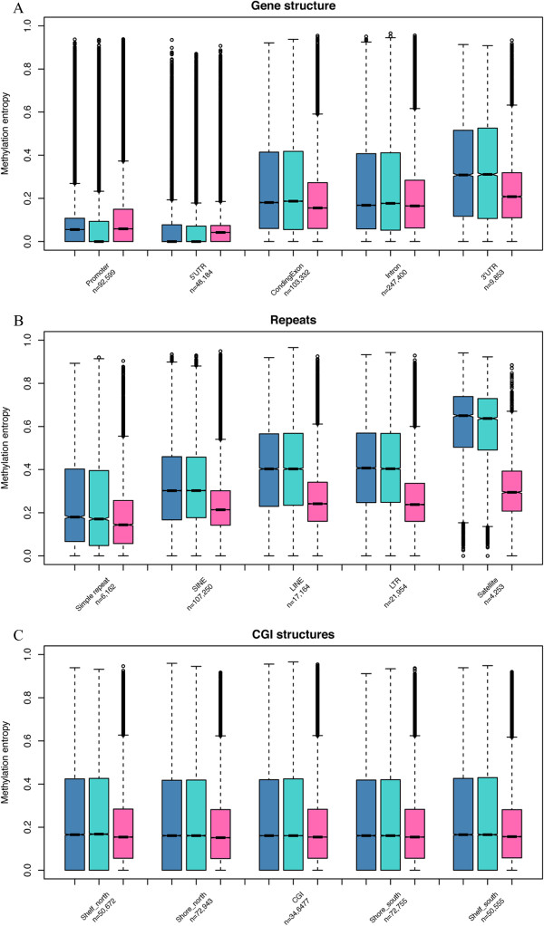
Figure 3.

DNA methylation entropy of different genomic regions for three methylomes. The ADS, ADS-adipose and ADS-iPSCs (from left to right) were plotted in each box plot where shows the median, upper and lower quartiles and 95% confidence intervals. The number of segments within each class was shown below the class label. (A) methylation pattern variation in gene structures. (B) methylation pattern variation in repeat regions. (C) methylation pattern variation in CpG island structures.
