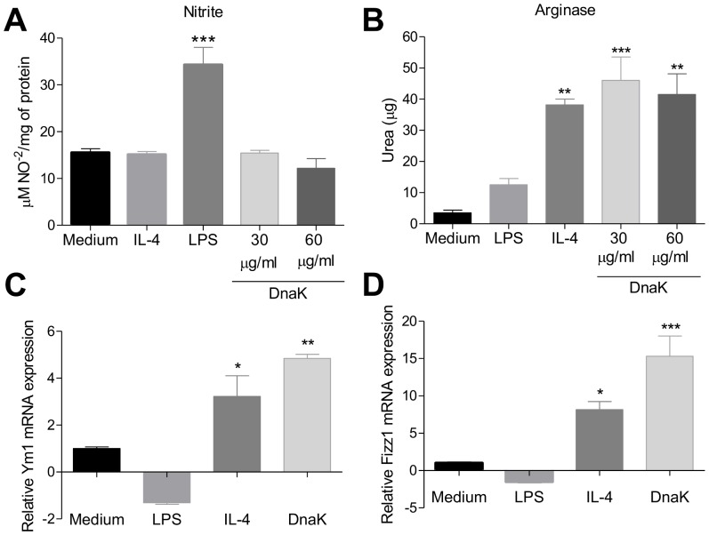
Figure 1. Extracellular DnaK induces the expression of M2 markers in bone marrow-derived macrophages.
BMMs were treated with LPS (30 ng/ml), IL-4 (40 ng/mL), DnaK (30 µg/mL or 60 µg/mL), or left unstimulated for 24 h. (A) iNOS activity was determinated by nitrite (NO2−) accumulation in the supernatant of macrophages. Data are the mean ± S.D. from triplicates. Data representative of three independent experiments. (***) p<0.001 indicates difference between LPS and other treatment groups. (B) Arginase activity was assessed by measuring the formation of urea from arginine. Data are the mean ± S.D. from triplicates. (**) p<0.01 and (***) p<0.001 indicate difference between treated groups and the medium group. Effect of DnaK on Ym1 (C) and FIZZ1 (D) expression in macrophages were quantified by real time PCR. The total amount of Ym1 and FIZZ1 mRNA were normalized to β-microglobulin signals and expressed as 2−Δ/ΔCT. The values represent means ± SEM from triplicates. Data representative of three independent experiments. All data were analyzed by one-way ANOVA with Tukey post hoc test.

