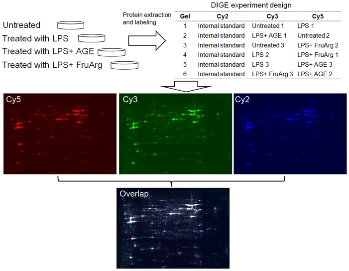
Figure 3. 2D-DIGE experimental design and workflow.
Lysates of BV-2 cells untreated or treated with LPS in the absence or presence of AGE (0.5%) or FruArg (3 mM) in triplicate were labeled with fluorescence CyDyes following the scheme shown in the table. The samples were then mixed and resolved on six independent 2D-DIGE gels. Three fluorescence images were obtained from each gel and subjected to image analysis using the SameSpots software.

