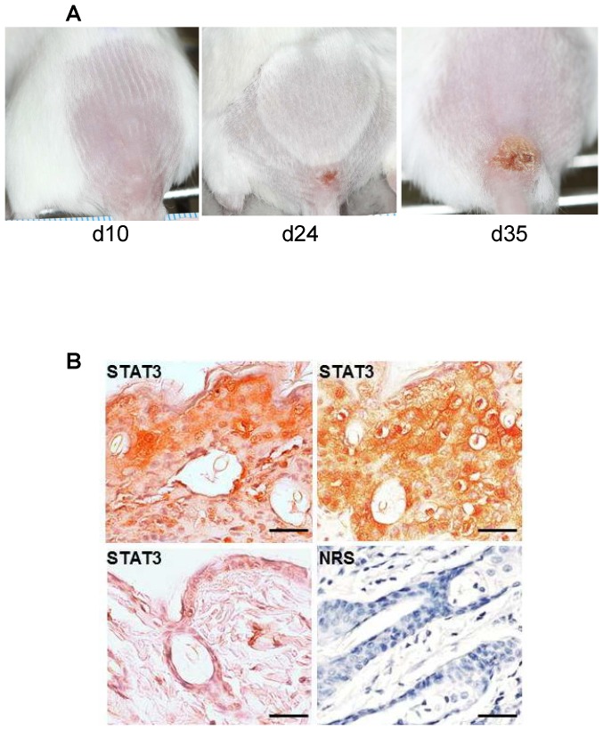
Figure 1. STAT3 detection in the primary vaccinia lesion of infected SCID mice.
A) Slowly growing primary lesion in vaccinia infected SCID mice. B) STAT3 detected by immunohistochemistry in formalin fixed and paraffin embedded skin tissue from terminal vaccinia lesions of infected SCID mice (top panels) or uninfected SCID mouse (lower left). Lower right panel, normal rabbit serum negative control antibody detection in representative terminal vaccinia lesion. Scale bars, 100 µm.

