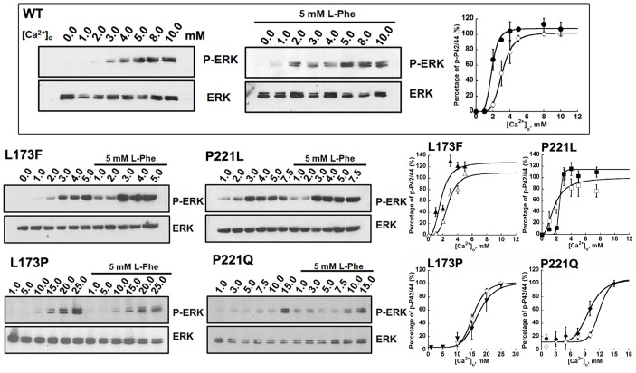
Figure 6. L-Phe potentiates [Ca2+]o-activated ERK signaling in CaSR-transfected HEK293 cells.
HEK-293 cells transfected with WT CaSR or its mutants were incubated in serum-free high glucose MEM medium containing 0.2% BSA overnight. Cells were washed with Hank's balance salt solution (HBSS) and then incubated in the presence of various Ca2+ concentrations (0.0-∼25.0mM) in the absence or presence of 5 mM L-phenylalanine for 10 min at 37°C. The incubations were stopped by exposure to the lysis buffer and processed for SDS/PAGE and Western blotting as described in the Methods. The Western blot results were further quantified using Image J. All [Ca2+]o-concentration response curves were normalized to the maximum response in each individual experiment. The Hill equation was employed to fit the data. Markers: [Ca2+]o only; closed markers: with L-Phe.

