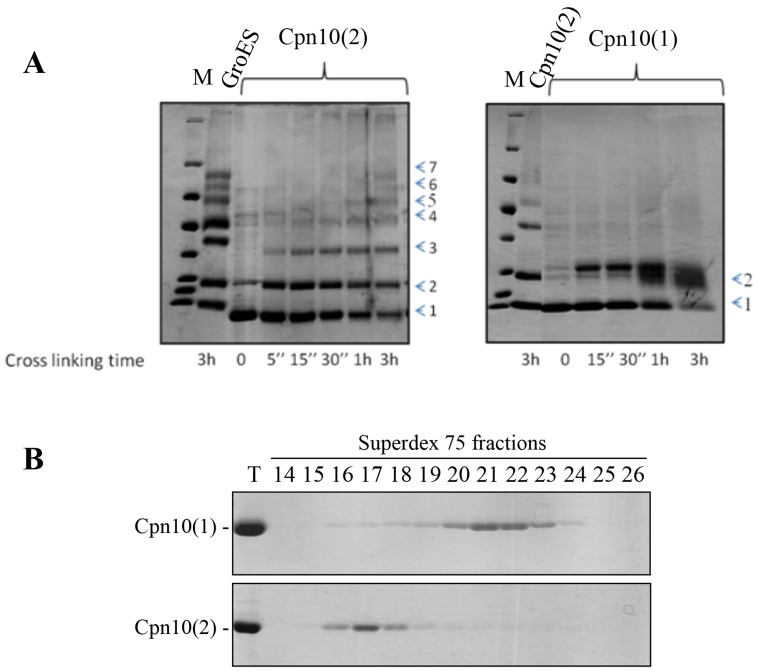
Figure 1. The oligomeric state of Cpn10(1).
(A) Time-dependent crosslinking pattern of Cpn10(1) (right panel) and Cpn10(2) (left panel). 45 µM of co-chaperonin was exposed to 1 mM DSS for the indicated times. The crosslinking products were separated on a mini gradient SDS acrylamide gels (10–19%), followed by staining with Coomassie blue. The numbers at the right indicate the number of cross-linked subunits in each species. (B) Elution profile of ∼1 mg Cpn10(1) (top panel) or ∼1 mg Cpn10(2) (lower panel) separated by gel filtration on a Superdex 75 column at room temperature at a flow rate of 1 mg/ml. Fractions were analyzed by SDS-PAGE (10 µl per lane). T = 5 µg of Cpn10. Fractions 15–18 contain primarily heptamer while fractions 20–23 contain primarily monomer - dimer.

