
Figure 4.
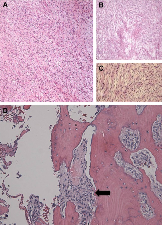
H&E microphotographs at (A and B) 4× and (C) 10× magnification, demonstrating moderately differentiated spindle cells with almost no mitoses. The hyperchromatic cells, which have coarse chromatin with mild pleomorphism, are arranged in short fascicles that split and merge, giving the classical herringbone architecture of fibrosarcoma. (D) Malignant spindle cells are seen in a fascicular pattern invading adjacent inferior orbital rim bone (black arrow).
Abbreviation: H&E, hematoxylin and eosin.