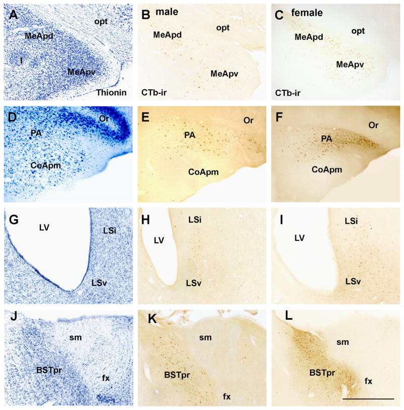
Fig. 5.
Distribution of retrogradely labeled neurons in the male and female forebrain. A, D, G, J Brightfield images of a reference brain stained with thionin showing the cytoarchitecture of a control (female) brain. B and C Brightfield images showing cholera toxin b immunoreactive neurons (CTb-ir) in the posterodorsal and posteroventral subdivisions of the medial nucleus of the amygdala (MeApd and MeApv, respectively) of male (B) and female (C) rats. E and F Brightfield images showing CTB-ir neurons in the posterior nucleus of the amygdala (PA) of male (E) and female (F) rats. H and I Brightfield images showing CTb-ir neurons in the lateral septum ventral (LSv) and lateral septum intermediate (LSi) of male (E) and female (F) rats. J and K Brightfield images showing CTb-ir neurons in the principal subdivision of the bed nucleus of the stria terminalis (BSTpr) of male (H) and female (I) rats. Abbreviations: CoApm, subdivision posteromedial of the cortical nucleus of the amygdala; fx, fornix; I, intercalate nucleus of the amygdala; LV, lateral ventricle; opt, optic tract; Or, stratus oriens; sm, stria medularis. Scale bar: 300 μm.

