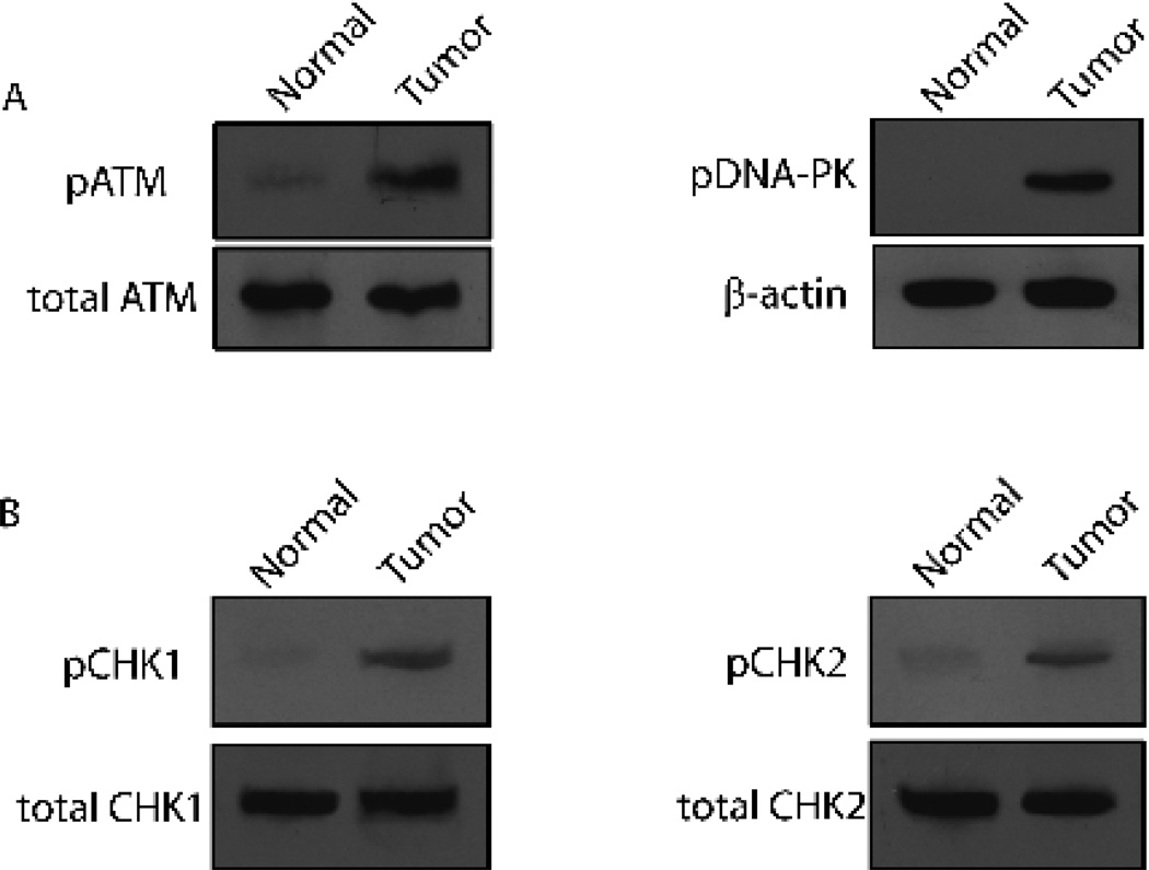
Figure 2. Checkpoint activation in human pancreatic cancer.
(A) Phosphorylation status of ATM and DNA-PK was tested by Western Blot from normal (C1-C3) and tumor (T2-T5) pancreatic tissues. Representative results from C3 and T5 are shown. Total ATM and β-actin were used for the loading controls, respectively. (B) Phosphorylation status of CHK1 and CHK2 was tested by Western Blot from normal (C1-C3) and tumor (T2-T5) pancreatic tissues. Representative results from C1 and T1 are shown. Total CHK1 and CHK2 were used for the loading controls, respectively.

