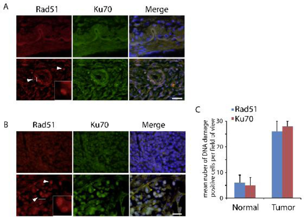
Figure 3. Activation of DNA repair pathways in human pancreatic cancer.
(A) Immunostaining of RAD51 and KU70 of duct was performed from normal (C1-C3) and tumor (T1, T2, and T4) pancreatic tissues. Representative images from C2 and T1 are shown. (B) Immunostaining of RAD51 and KU70 of pancreatic body was performed from normal (C1-C3) and tumor (T1-T4) pancreatic tissues. Representative images from C2 and T3 are shown. Enlarged box denotes RAD51 foci. Scale bar: 20 µm. (C) Numbers of positive Rad51 and Ku70 cells around the duct in normal pancreas (C1-C3) and pancreatic tumor (T1, T2, and T4) were counted. Three independent experiments were averaged. The error bars represent the SD.

