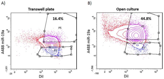
Figure 7. FACS analysis of cells grown in open culture vs. separated by membrane filter in modified invasion chambers.

Adapting our model of in vitro cell culture of osteosarcoma cells and osteoblast cells, we used modified Boyden chambers including membrane filters with the smallest commercially available pore-size (0.4 μm, or 400 nm). At 48 hours, cells in the bottom chambers were collected and sorted using fluorescence-activated cell sorting (FACS); this step separated out single-positive non-recipient cells that were excluded from further analysis. (A) FACS analysis of double-positive MC3T3 cells following transwell assay for 48 hours, versus (B) FACS analysis of double-positive MC3T3 cells in open co-culture with K7M2 for 48 hours (without membrane barrier separation). The difference in these results was statistically significant with a p-value of 0.0002; the standard deviation was 2.15 for the transwell plate and 3.50 for the open culture experiments.
