
Fig. 3.
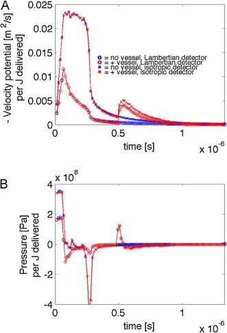
Velocity potential (−ϕ) and pressure (P) per J delivered to the skin with 500-μm-dia. blood vessel centered at 1 mm depth below the skin surface. (A) The time courses of the magnitude of velocity potential, −ϕ(t), for four cases, skin ± a vessel and detector collection either isotropic or Lambertian (detector flat on skin surface). (B) The pressure P(t) for the four cases. After the initial response to the superficial deposition of energy in the pigmented epidermis (370 Pa) and the blood-perfused dermis, the delayed signal from the vessel arrives at 0.5 μs. The isotropic detector is sensitive to the epidermal melanin, and there is a strong negative pressure due to the edge of the 0.8-mm-dia. beam of irradiation. The Lambertian detector is insensitive to this edge effect in the epidermis. The vessel pressure signal is the same for both isotropic and Lambertian detectors.