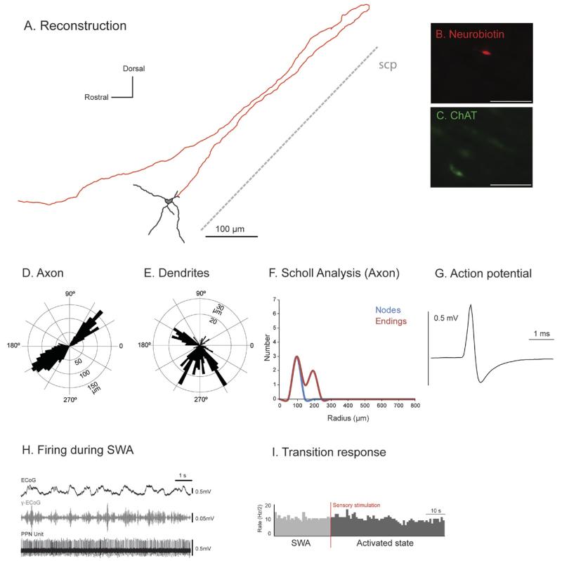
Fig. 3. Tonic non-cholinergic PPN neurons do not modify their activity during transitions from slow oscillations to global activation.
(A) Reconstruction of the cell body, dendrites (black) and axon (red) of an individual tonic non-cholinergic PPN neuron. These neurons typically gave rise to an axon that emerged caudally from a secondary dendrite, formed a “loop” and traveled rostrally giving rise to an ascending axonal projection. (B, C) PPN neurons were juxtacellularly labeled and identified as non-cholinergic by the lack of co-localization of fluorescent markers for neurobiotin (B) and ChAT immunoreactivity (C). (D, E) Polar histograms show direction of arborizations in the sagittal plane of the axon (D) and dendrites (E). (F) Scholl analysis shows the number of nodes and endings relative to the distance (in μm) from the soma. (G) Average action potential shape (0.78 ms width). (H) During robust SWA, neocortical activity (ECoG; black) was dominated by a slow oscillation (~1 Hz), the active components of which supported nested gamma oscillations (30–50 Hz; γ-ECoG, grey). This non-cholinergic neuron fired at a high frequency (22 Hz) and did not show any preferential firing during the phases of the neocortical slow oscillation (top trace). (I) FR of the same tonic firing neuron during the transition from neocortical slow oscillations to an activated state (indicated by the vertical bar; sensory stimulation, red), showing that tonic neurons do not significantly change their FR (three of four). Scale bars: 100 μm.

