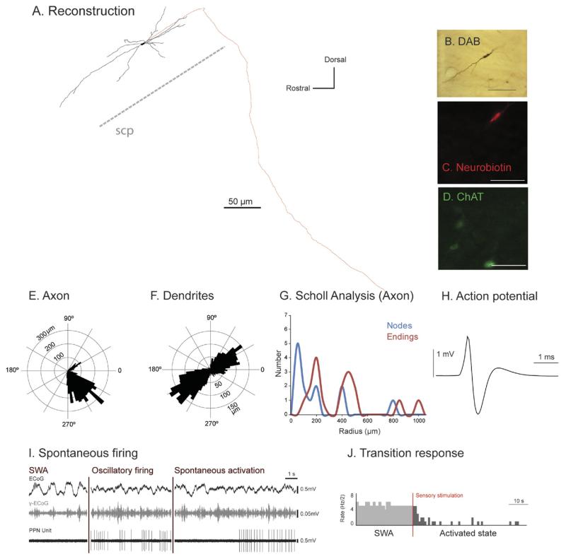
Fig. 5. Irregular firing descending-axon non-cholinergic PPN neurons show variable patterns of activity across different brain states.
(A) Reconstruction of the cell body, dendrites (black) and axon (red) of an individual irregular firing non-cholinergic PPN neuron. These neurons typically gave rise to an axon that emerged caudally from a primary dendrite; this neuron had a descending-only projection and did not give rise to any local boutons within the PPN. (B–D) PPN neurons were juxtacellularly-labeled (B) and identified as non-cholinergic by the lack of co-localization of fluorescent markers for neurobiotin (C) and ChAT immunoreactivity (D). (E, F) Polar histograms show direction of arborizations in the sagittal plane of the axon (E) and dendrites (F). (G) Scholl analysis shows the number of nodes and endings relative to the distance (in μm) from the soma. (H) Average action potential shape (0.72 ms width). (I) During robust slow-wave activity, neocortical activity (ECoG; black) was dominated by a slow oscillation (~1 Hz), the active components of which supported nested gamma oscillations (30–50 Hz; γ-ECoG, grey). This non-cholinergic neuron showed spontaneous periods of silence alternated with stereotyped activity and showed no clear temporal relationship to the neocortical slow oscillation (top trace). (J) Example of induced cortical activation of the same PPN non-cholinergic neuron during the transition from neocortical slow oscillations to an activated state. Shortly after the onset of a hindpaw pinch this neuron was inhibited. Scale bars in (B) 50 μm, and (C, D) 20 μm.

